How Gemelli Hospital uses AI to streamline prostate cancer workflows

Gemelli Hospital (Source: Vatican News)
Gemelli Hospital in Rome is one of Italy’s largest academic medical centers, known for its focus on clinical care, research, and education. Within its radiology department, the team has adopted Lucida Medical’s AI solution, Prostate Intelligence (Pi™), to support prostate MRI interpretation and improve radiology workflow in prostate cancer diagnostics.
This case study outlines how AI has been integrated into daily practice, helping clinicians manage reporting tasks, prioritise cases more effectively, and better collaborate with urology teams. The experience at Gemelli Hospital reflects a broader shift toward AI-driven workflow optimization in prostate cancer care.
Identifying workflow challenges
Dr. Luca Russo and his colleagues began exploring AI tools to address specific challenges in prostate MRI. They needed a solution that could integrate with their PACS system, work within their existing workstation environment, and support consistent, high-quality reporting. The goal was to improve diagnostic confidence, reduce time spent on repetitive tasks, and support decision-making, especially in complex or borderline cases.
One of the most time-consuming parts of the workflow was calculating prostate volume manually using the ellipsoid formula. This process could take several minutes and was not always accurate, especially in patients with irregular anatomy. In addition, managing PI-RADS 3 cases was difficult since these borderline lesions often led to uncertainty and inconsistent decisions between readers.
How Pi™ supports the clinical workflow
Pi™ was integrated into the hospital’s diagnostic pathway, including acquisition, prioritisation, analysis, and reporting. It helps pre-analyse prostate cases, and sorts them into likely positive or likely negative. The prioritisation allows doctors to focus on what matters most, based on urgency or complexity. This flexibility is useful throughout the day, especially when fatigue affects concentration.
“Now, I start my day knowing which cases are likely negative or positive. The prioritisation function is something I couldn’t imagine practicing without,” says Dr. Russo.
Beyond prioritisation, Pi™ also assists with decision-making. Radiologists use it to reaffirm their assessments, particularly in borderline or complex cases, which contributes to greater diagnostic confidence and consistency across the team.
Volume calculation is now automatic and more precise. Pi™’s segmentation accounts for anatomical variations, such as post-TUPR changes or large BPH nodules, which improves PSA density estimates and biopsy decisions. According to Dr. Russo, integrating Pi™ into the clinical workflow has made a real difference:
“Tasks like volume calculation used to slow us down, but now we’re faster and more confident in our diagnoses, especially the less experienced radiologists.”
Supporting biopsy decisions with AI
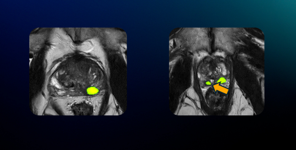
Lucida Medical’s Pi™ has helped clinicians at Gemelli Hospital make more informed biopsy decisions by highlighting features that may indicate benign or clinically significant disease.
In one case, Pi™ detected a smaller lesion that had not been initially noticed. Both lesions were later confirmed by biopsy as clinically significant cancers, supporting the value of AI-assisted review in complex cases.
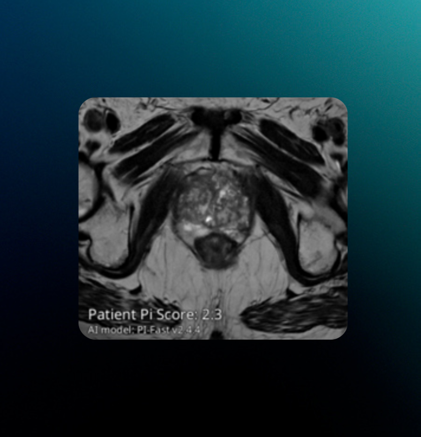
In another case, a patient referred with a PI-RADS 4 lesion underwent MRI at Gemelli Hospital. Pi™ did not identify a focal lesion, and further review of high-quality T2-weighted images revealed the nodule to be consistent with ectopic BPH. Based on this assessment, the team decided not to proceed with biopsy. Pi™’s output aligned with the radiologist’s interpretation and contributed to a more confident decision to avoid unnecessary intervention.
Streamlined and more targeted biopsy planning
Lucida Medical’s Pi™ supports coordination between radiology and urology teams by providing structured reports that include heatmaps and biopsy segmentation. These visuals help guide biopsy planning and reduce the time required for manual prostate contouring, especially in complex cases.
By streamlining image interpretation and biopsy preparation, Pi™ contributes to a more efficient workflow across departments. Radiologists can deliver clearer guidance, and urologists benefit from faster, more targeted planning. This integration supports a smoother diagnostic pathway for prostate cancer and strengthens communication between specialties.
Advancing the prostate cancer pathway with AI
Looking ahead, Dr. Russo anticipates that AI tools like Pi™ will become more accurate and more deeply integrated into clinical workflows. He sees potential for AI to support the entire prostate cancer pathway, from initial MRI interpretation to biopsy planning, and eventually histological analysis with diagnostic scoring.
This progression could help standardise decision-making, reduce variability across specialties, and improve coordination throughout the diagnostic process. As AI continues to evolve, its role in supporting prostate cancer care is expected to expand beyond imaging into broader clinical applications.
If you are exploring ways to streamline prostate cancer diagnostics and improve workflow efficiency, Lucida Medical can help. To learn more about how Pi™ supports clinical decision-making and pathway coordination, get in touch with us at contact-us@lucidamedical.com or request a demo through our website.